AI Image Analysis
Offering machine learning image processing and analytics for all imaging modalities. Transform your images into drug development insights.

Digital Transformation with Image Segmentation
At digiM, we specialize in transforming complex imaging data into actionable insights. Whether you're working with micro-CT, SEM, optical microscopy, or polarized light microscopy, our advanced image analysis workflows are built to extract critical structural, spatial, and quantitative information from your data. Backed by decades of experience in pharmaceutical, materials science, and life science applications, we support clients by delivering high-quality, filing-ready results and enabling deeper understanding through imaging. In addition to drug products, we support analysis of tissue and clinical imaging data, such as MRI and CT, enabling anatomically accurate meshes for drug delivery modeling.
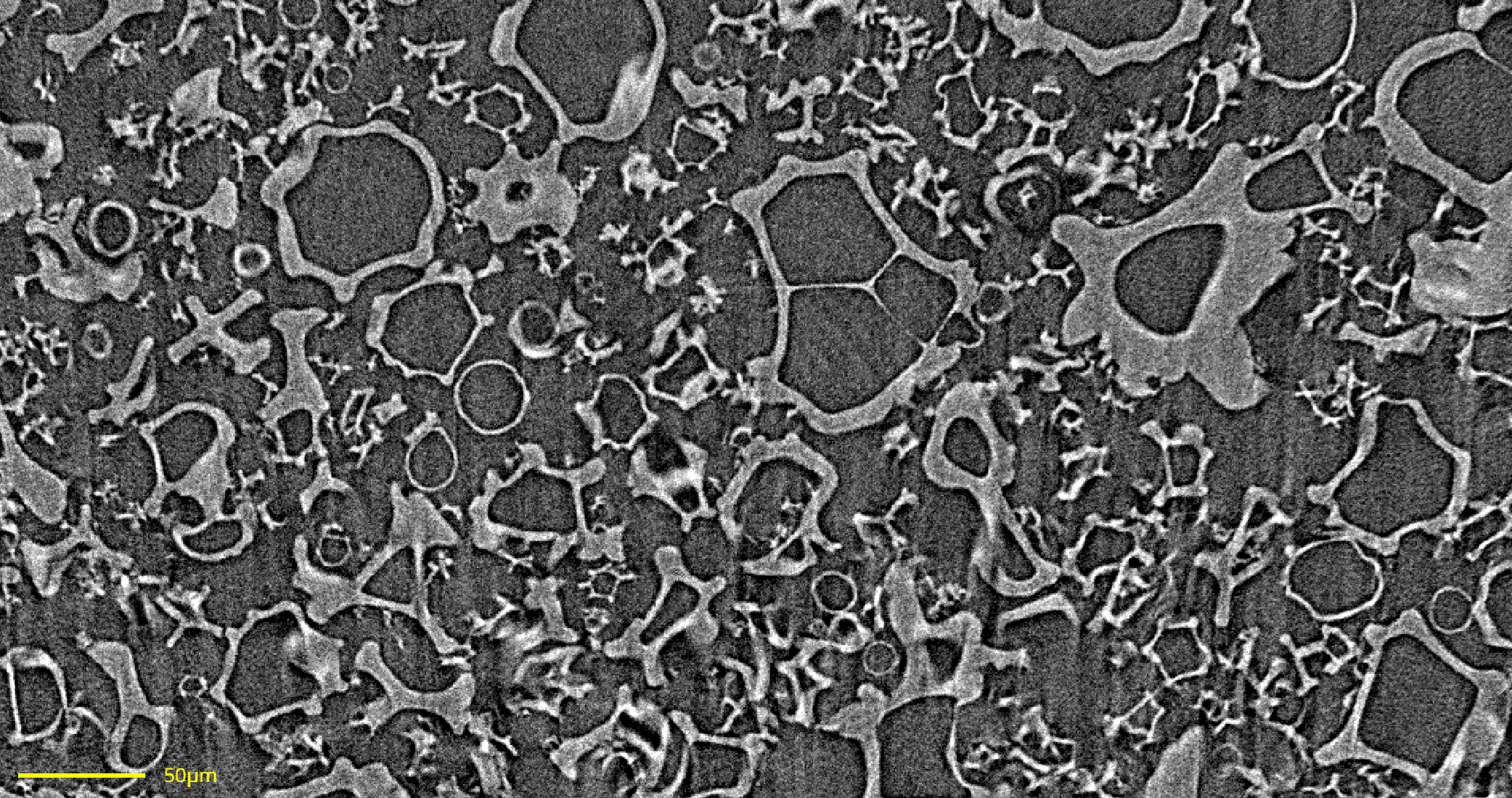

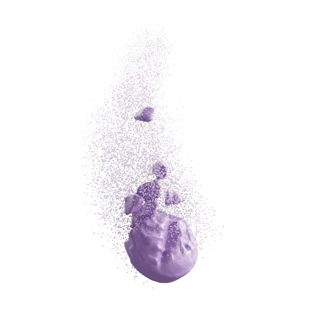
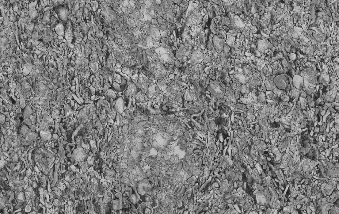
Image segmentation is the foundation of our analysis — classifying meaningful features to quantify components, structures, and regions of interest. Using our custom tools, we segment complex images into clean, analyzable parts with precision. The before-and-after slider below shows how we isolate key features in an X-ray micro-CT scan of a spray dried dispersion, revealing detailed analysis of properties like pore size, wall thickness, and particle morphology.
Thresholding
Machine Learning
Deep Learning
Fast and effective for high-contrast data, thresholding offers simple yet powerful classification based on grayscale values.
Our supervised machine learning methods leverage expert-labeled data to deliver accurate, reproducible segmentations across diverse sample types.
For the most complex images, our deep learning models handle low-contrast, noisy, or irregular data, achieving human-like precision.
From Image to Quantification
Powered by digiM I2S, our analytical services can help transform your real world sample into a digital dataset with quantifiable insights. These digital twins support research and development through a detailed understanding of microstructure and drug product quality. Direct from the imaging data, we extract important attributes which are difficult or impossible to extract with routine analytical measurements.
Common quantifications include:
- Content uniformity and spatial distribution
- Coating thickness
- Particle size, surface area, and shape
- Ingredient specific porosity analysis
- Particle surface area

Let’s Analyze Your Images
Looking to turn your imaging data into insights? digiM offers image analysis as a service—just send us your scans, and we’ll take care of the rest. Our team works as an extension of your lab, delivering rapid, customized analysis with full documentation and support. Whether it’s a single study or ongoing projects, we’re ready to help you unlock the full potential of your images. Contact us today to get started.

Transform Your Program with Microstructure Science
Get started with a drug product digital twin.