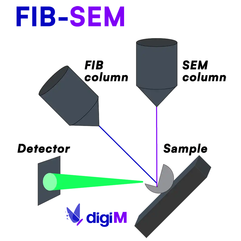
FIB-SEM
Focused Ion Beam Scanning Electron Microscopy is a technique for nanometer scale 3D imaging, typically targeting 3 to 50 nm resolution.

FIB-SEM analysis for drug product development
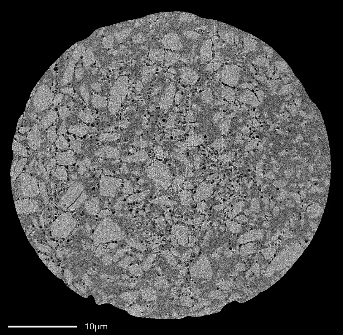
For pharmaceutical drug product development, FIB-SEM analysis is predominantly used in the evaluation of porosity and particle networks at the nanoscale. Common applications include complex controlled release dosage forms like microspheres, implants, and IUDs. It has also been applied to evaluate beads, coatings with pore formers, and in situ formed depots. Combining 3D FIB-SEM and AI analytics enables quantification of API domain and porosity attributes within the final drug product. These measurements are difficult, if not impossible, to capture with other methods. With these data, we often support the construction of data packages for ANDA and NDA filings.
Upon digital reconstruction of 3D FIB-SEM cubes, the API particle and porosity networks can be used for image-based simulation of mass transport phenomena. For controlled release products, this includes a direct prediction in situ of drug release profiles.

What is FIB-SEM?
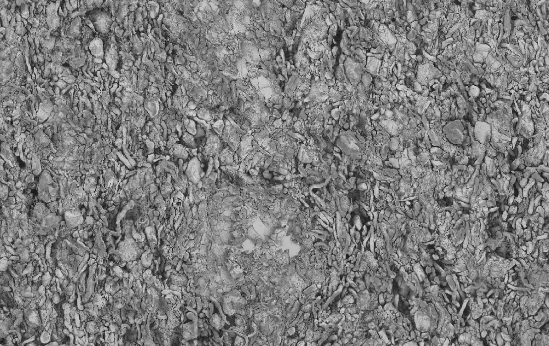
Compared to conventional SEM imaging which is limited to surface investigations, FIB-SEM adds a “milling” tool in the form of a focused ion beam (FIB). Due to its heavy ionic mass, the FIB ions remove a small amount of material in a sputtering (or milling) process. FIB thus exposes the structures below the surface for direct visualization with SEM. Unlike conventional cross-sectioning methods which can leave rough, uneven surfaces, FIB removes layers as thin as 3nm, providing a flat cross-section with minimal surface defects. Repeated FIB milling followed by consecutive SEM imaging produces a stack of images that are reconstructed into a 3D volume.
End-to-end FIB-SEM analysis services
While FIB-SEM is a destructive technique, it is critical that the inherent microstructures are not destroyed during data collection. This is especially true for soft, low glass transition temperature materials, where a poor selection of imaging and milling parameters can create artifacts and obscure the real structures within. From decades of combined experience we have mastered the use of FIB-SEM analysis, ensuring that microstructures are preserved and collected accurately. Where 3D FIB-SEM volume collection might take some labs months of planning and optimization, we routinely perform 3D FIB runs overnight.
To account for sample size and material properties, we offer the entire suite of FIB techniques including cryo-FIB SEM, gallium ion beam sources, and argon ion beam sources. FIB data collection can also be combined with EDX spectroscopic mapping. For larger samples, cryo-microtome sectioning can be performed in preparation for FIB-SEM collection. Our FIB-SEM services include the whole suite of analysis, from dataset collection to image processing, structure analytics, and modeling.

Our Expertise In Numbers

Be Part of The Therapeutics Revolution
Revolutionize drug development with our AI-driven platform. Get started today.