Mosaic SEM
Mosaic SEM is an imaging technique using SEM resolution (5-200 nm) to cover a large field of view that is mm to cm in size.

What is Mosaic SEM?
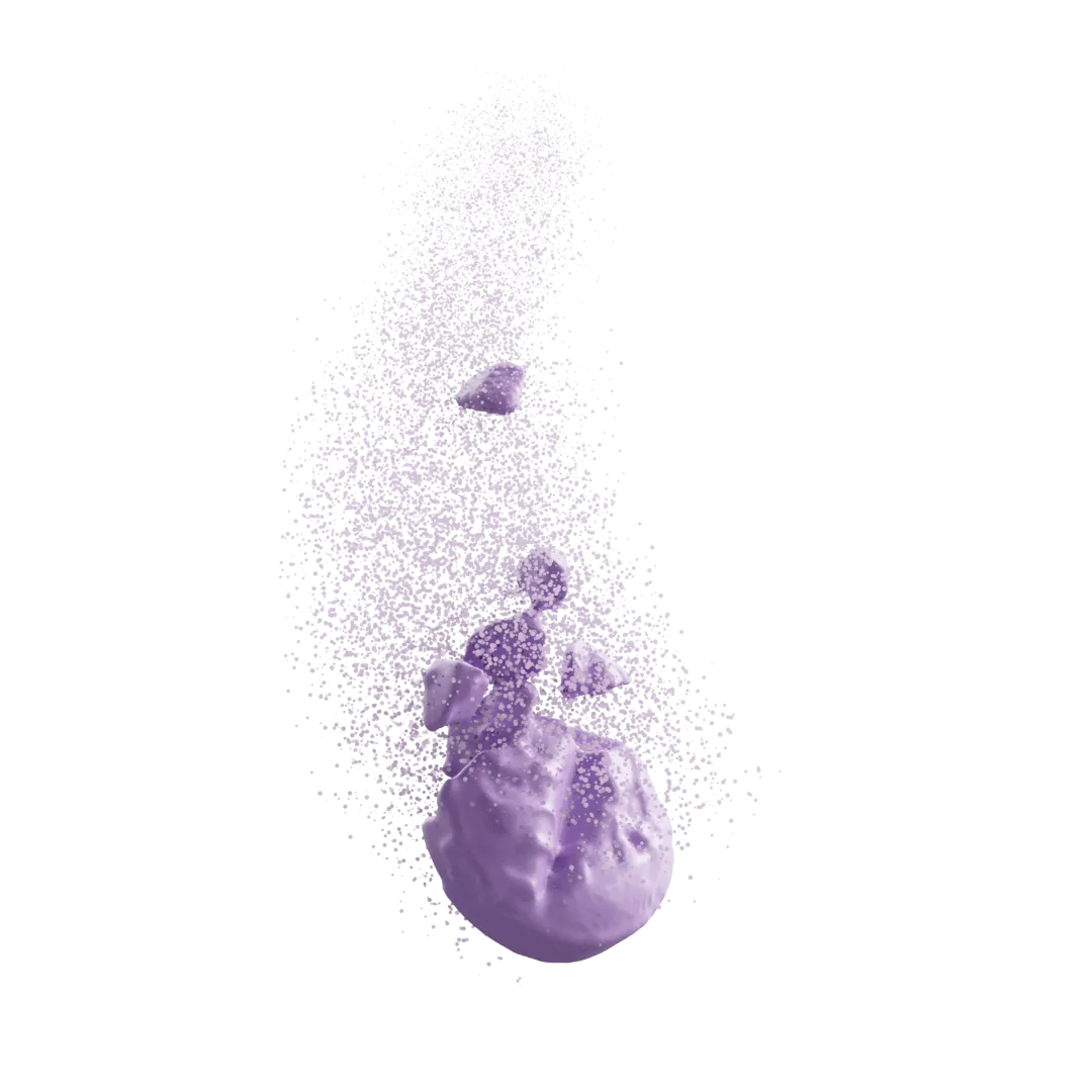
Mosaic SEM is a state of the art workflow combining specially prepared cross-sections with a high-resolution map of stitched images. Compared to conventional SEM imaging which is limited to surface investigations, Mosaic SEM enables a direct visualization of internal structures across a large area. The process involves a combination of cutting and polishing steps with an argon ion beam to expose a sample cross-section. After polishing, hundreds of images collected at nanometer resolution are stitched together to create a “mosaic” map of interior structures with a field of view covering millimeters. Similar to FIB-SEM, Mosaic SEM provides a fine level of detail which is not readily visible from the exterior view of the sample. With its large area coverage, Mosaic SEM bridges the field of view gap between localized FIB-SEM analysis and full sample 3D X-ray micro-CT analysis.

End-to-end Mosaic SEM analysis services
While Mosaic SEM is a destructive technique, it is critical that the inherent microstructures are not destroyed during data collection. This is especially true for soft, low Tg materials, where a poor selection of imaging and milling parameters can create artifacts and obscure the real structures within. From decades of combined experience we have mastered a complete workflow for Mosaic SEM analysis, ensuring that microstructures are preserved and visualized accurately. Where high-resolution cross-section studies might take some labs months of planning and optimization, we routinely perform Mosaic SEM map collection in a few hours. We design each Mosaic SEM experiment carefully, optimizing the cross-section preparation conditions to account for the sample size and material properties. Cryo-microtome sectioning can be applied as a preparation step for the Mosaic SEM experiment, and data collection can be combined with EDX spectroscopic mapping. Our Mosaic SEM services include the whole suite of analysis, from dataset collection to image processing, structure analytics, and modeling.

Mosaic SEM analysis for drug product development
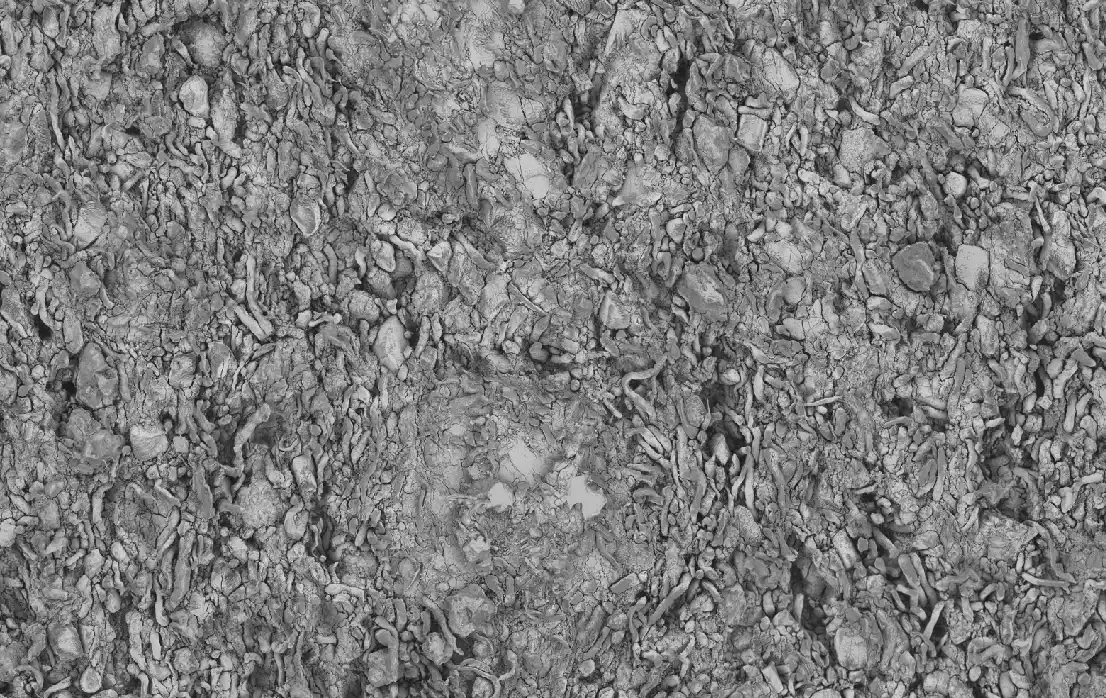
For pharmaceutical drug product development, Mosaic SEM analysis is often used to evaluate the interaction of ingredients at the nanoscale. Common applications include tablets, implants, coatings, and thin films. This analysis enables direct quantification of API particles and porosity attributes within the final drug product. These measurements are difficult, if not impossible, to capture with traditional analytical methods. For tablets, Mosaic SEM can help assess ingredient interactions, compaction behavior, coating-core interfaces, disintegrant functionality, and root causes of fracture formations.


Formulations
Studied

Be Part of The Therapeutics Revolution
Revolutionize drug development with our AI-driven platform. Get started today.