Shawn Zhang, Ph.D.
Co-founder, Managing Director
Explore how image-based methods outperform MICP in measuring porosity.
Let's take a minute to consider porosity and how it's measured.
At the simplest level, porosity can be derived using principles like solid fraction, given that the material's true densities are known. Pharmaceutical products, however, introduce challenges to volume and density measurements. The volume of a compacted tablet will expand after ejected from the die and create porosity or fractures along the way. The multicomponent nature of various ingredients in the tablet formulation complicates density estimation. Volume measurements for particulate samples, either as final drug products such as long acting injectable (LAI) microspheres or as intermediates such as mixes from dry granulation, are even more difficult. These particles are irregular in shape, often porous, and light. We can measure the volume of a bed of particles, but that is far different from the actual volume.
Liquid intrusion and gas adsorption are two additional methods widely adopted in the industry to measure porosity. Other than the limitation that such external fluid penetration method cannot measure closed pores, there is reasonable success in measuring porosity for rigid materials such as rocks, bone, and ceramics.
However, when measuring porosity of soft pharmaceutical materials, such physical measurements can go awry.
Figure 1 below shows porosity measurements for several long-acting microspheres, comparing the outputs from mercury injection capillary pressure (MICP) and an image-based method (FIB-SEM and AI image processing). The difference is clear as day. MICP has significantly higher pore volume, and has no discriminatory power between these microsphere batches. With the image-based method, we can identify these differences.

Why are these measurements so different? To compare these in a truly quantitative way, we can perform a digital mercury porosimetry simulation on a real sample's imaging data for a 1:1 comparison with the lab MICP results.
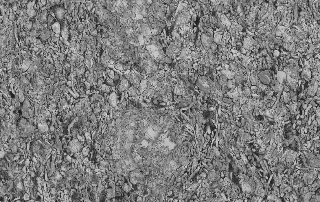
Let's start by taking a look at the digital MICP applied to an X-ray microscopy scan of a microsphere powder bed, the gray curve in the figure 2 below. The region 1, shows excellent overlap with the lab-based MICP, providing a validation on the simulation and identifying what type of porosity is being measured from the mercury intrusion. Namely, this measurement was reading the air space between each sphere in the powder bed.

What about smaller pores? Region 2 covers pores in the range of 0.01-1 micron, where pores inside the spheres are expected to fall. This digital MICP experiment was run from the real-world pore network identified from 3D FIB-SEM analysis. The visual based assessment of porosity provides evaluation of all pores, regardless of their connection to each other or the external surface. The digital MICP will only quantify those pores which are connected, which in this case is a fairly strong network at higher volume %. How about lab-based MICP? In this region the measurement is almost entirely flat, not picking up on this strong porosity network. Traditional MICP failed to measure porosity in this region because there were little to no surface pores connected to the inner network.
Lastly, we have the tail of the lab-based MICP curve, where there is a spike in measured pore fraction, with pores in a size range not identified from imaging. This phenomenon is an area of continued research - it is suspected that this tail includes artificially induced porosity from the excessive pressures applied to a soft material matrix.
✅ Digital based MICP has been validated directly against lab-based MICP.
✅ Digital MICP can be used applied to imaging data at various scales, capturing the entire family of porosity types.
❌ Lab-based MICP failed to pick up on the porosity network of the microspheres, due to no connection to the interior porosity.
❌ Lab-based MICP needs to be approached very carefully when evaluating soft pharmaceutical materials, which are prone to deformation and may introduce artificial pore readings.
✅ Image-based porosity measurements provide a more direct and accurate pathway to quantify the complexity and connectivity of different pore types, including subtleties that traditional methodologies may miss.


Get started with a drug product digital twin.