Synchrotron micro-CT Imaging: “You mustn’t be afraid to dream a little bigger, darling”
What is Synchrotron micro-CT imaging? Dreaming bigger for superior resolution, collection times, and data quality


I don’t know about you, but Christopher Nolan’s movie Inception is one of my favorite movies, and I must have seen it at least four times in theaters. Every time a new detail jumped out at me, and the whole picture started to click more firmly in place, and of course the sharp quick wit of the characters never lost its appeal. Tom Hardy’s comment to Joseph Gordon-Levitt “You mustn’t be afraid to dream a little bigger, darling,” always tickled me. He’s absolutely right: to really achieve our goals, we always need to dream bigger…and bigger is always better. This also rings true for micro-CT imaging, and I don’t mean bigger images.
Standard lab-based micro-CT systems range in size from benchtop instruments designed to fit on a lab bench in any laboratory, to large cabinet systems weighing in at 1,500 kg. The benchtop systems are wonderful tools, capable of capturing quick visualizations and some longer time scans with good resolution. They’re the jack-of-all-trades micro-CT and in many cases are all that a small characterization lab needs for its X-ray imaging. The trade off here is the sample size, X-ray source, and max magnification are constrained by needing to fit into such a compact space. For characterization needing higher resolution images or needing specialized detection methods and X-ray characterization, the larger lab-based systems are needed. Systems like the Zeiss Versa Xradia series can perform local tomography with clever optical magnification to achieve 500 nm/pixel resolution with relatively large samples. Other large systems like the Rigaku nano3DX can achieve similar resolutions but also offer quasi-monochromatic X-ray imaging with their variable X-ray source containing copper, chromium, and molybdenum. Naturally these systems require a larger lab space due to their footprint, but once installed they can regularly output extremely high-quality images that offer incredible insight into a system.
All that said, there’s a major Achilles’ heel for all lab-based micro-CT systems: focusing the X-rays. For visible spectrum light, you can magnify the light by using lenses to focus the light onto a single point. That’s how traditional microscopes work: using different lenses to magnify the light that goes through a sample. X-rays don’t really like to be focused. There’s no single material that can readily bend X-rays since the photons will just pass right through them. There are strategies to focus X-rays, like grazing incidence reflection systems, and capillary condenser tubes but those drastically reduce the X-ray beam intensity and worsen the image quality. So, unless your X-ray beam is thousands of times brighter and more intense than conventional lab sources, these options aren’t feasible. But once again, we have Tom Hardy whispering into our thoughts: dare we dream even bigger.

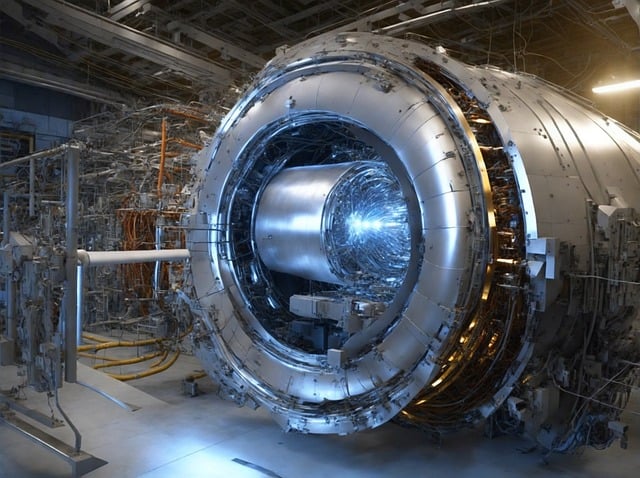
This is where we turn to particle physics. Particle accelerators, known as synchrotrons, take charged particles like electrons or protons and accelerate them to very high velocities. These particles whirl in circles where they are kept on path by electromagnets that bend them into a circle. However, bending the particles into a circular path changes the particles momentum, and because we know momentum is conserved that leftover momentum must go somewhere. That spare momentum gets converted to energy and is emitted as light across the whole electromagnetic spectrum from radio waves up to X-rays. And here’s where it gets fun: the more momentum change there is, the larger the intensity of emitted light. So, if the particles are moving REALLY fast then the emitted light from the synchrotron is thousands to millions of times brighter than what is produced at a normal lab! So how fast are we talking here? At the National Synchrotron Light Source 2 (NSLS-II) at Brookhaven National Lab, the electrons are accelerated to 3 GeV, which means they are travelling at 99.99999885% the speed of light. I’d say we are definitely dreaming bigger now modern synchrotrons are referred to as third generation synchrotrons and are widely used across the world for a host of characterizations due to their ability to produce such intense light across the electromagnetic spectrum. Ultra precise X-ray scattering measurements, infrared spectroscopy that is spatially correlated, X-ray spectroscopy measurements, and most importantly for us computed tomography imaging. The X-rays at synchrotrons for CT imaging are both focused spatially to achieve resolutions as high as 20 nm/pixel as well as spectrally focused to achieve truly monochromatic X-ray beams. Even better, the X-ray beam is perfectly parallel allowing for faster reconstruction of the images, easier sample preparation, and the implementation of advanced phase-based imaging.
So how big are synchrotrons? Well if we look at NSLS-II again, the primary accelerator ring has a circumference of 792 meters, and has at least 3,100 tons of steel. While building one for your lab isn’t on the table, these facilities do offer beamline access to researchers requiring superior resolution and advanced image-based studies. We are fortunate to have these partnerships in place with national and international synchrotrons, bringing the best in data quality and advanced imaging to our clients. No longer a dream, bigger and better for X-ray CT is truly here, all with a “little” thanks to particle physics.
More Blog Articles
Transform Your Program with Microstructure Science
Get started with a drug product digital twin.


